May 23, 2025
by Mikhail Elyashberg, Leading Researcher, ACD/Labs
Computer Assisted Structure Elucidation of Mantidisflavin A Using Structure Elucidator Suite
Cheng and coworkers have developed an interest in searching for non-peptide small molecules from insects. They have characterized a series of structurally intriguing and biologically active molecules, with novel ring formations on usual skeletons from several insects. Recently the group focused on finding small molecules from the egg cases of the insect Tenodera sinensis. This investigation resulted in the isolation of an unprecedented 6/6/6/5/5 skeleton, mantidisflavin A (1), originating from riboflavin [1].
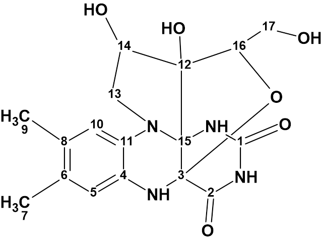
1
In this compound the riboflavin skeleton was supplemented by the formation of two additional fused heterocycles through a novel C−C bond and an oxygen bridge on the riboflavin backbone. The structure of mantidisflavin A was elucidated using 1D and 2D NMR spectra in combination with HR-ESI-MS and IR.
Mantidisflavin A was isolated as a white powder with molecular formula C17H20N4O6 (10 degrees of unsaturation), as determined by its 13C NMR and DEPT spectra as well as the negative HR-ESI-MS data at m/z 375.1302 [M −H]− (Calcd for C17H19N4O6, 375.1310).
The molecular formula and NMR data of mantidisflavin A (Table 1) were used to challenge Structure Elucidator Suite.
Table 1. NMR spectroscopic data of mantidisflavin A.
| C/X Label | δC | δcalc (HOSE) | XHn | δH | H Mult. | COSY | H to C HMBC |
| C 1 | 151.2 | 152.79 | C | ||||
| C 2 | 168.1 | 166.87 | C | ||||
| C 3 | 81 | 91.76 | C | ||||
| C 4 | 126.9 | 127.78 | C | ||||
| C 5 | 115.6 | 117.45 | CH | 6.5 | s | C 8, C 11 | |
| C 6 | 126 | 132.96 | C | ||||
| C 7 | 18.7 | 18.52 | CH3 | 2.02 | s | C 5, C 8, C 6 | |
| C 8 | 125.4 | 124.1 | C | ||||
| C 9 | 18.9 | 19.23 | CH3 | 2.08 | s | C 10, C 8, C 6 | |
| C 10 | 114.5 | 117.06 | CH | 6.34 | s | C 6, C 4 | |
| C 11 | 128.9 | 131.15 | C | ||||
| C 12 | 78.3 | 88.35 | C | ||||
| C 13 | 52.8 | 53.88 | CH2 | 2.74 | u | 4.43 | C 12, C 11 |
| C 13 | 52.8 | 53.88 | CH2 | 3.95 | u | ||
| C 14 | 71.7 | 74.86 | CH | 4.43 | u | 2.74, 5.52 | |
| C 15 | 85.5 | 82.74 | C | ||||
| C 16 | 80.2 | 84.34 | CH | 3.91 | u | 3.39 | C 14, C 3 |
| C 17 | 61.9 | 61.22 | CH2 | 3.39 | u | 3.91, 4.41 | |
| C 17 | 61.9 | 61.22 | CH2 | 3.51 | u | ||
| N 1 | NH | 7.69 | u | C 3, C 1 | |||
| N 2 | NH | 10.16 | u | C 3, C 2 | |||
| N 3 | NH | 6.67 | u | C 12, C 15, C 5, C 11, C 2 | |||
| O 1 | OH | 5.52 | u | 4.43 | |||
| O 2 | OH | 6.02 | u | C 14, C 12, C 16, C 15 | |||
| O 3 | OH | 4.41 | u | 3.39 |
The molecular formula of compound 1 shows that the molecule has a hydrogen deficiency. The ratio of the number of hydrogen atoms to the number of skeletal atoms is 0.7, which, according to Crew’s rule, makes the structure elucidation of this unprecedented compound a challenging problem. As it often happens now, unfortunately, the article does not contain a table of all HMBC correlations. Instead, only the key HMBC and COSY correlations are presented graphically (Figure 1).

Figure 1. Key HMBC and COSY correlations of 1.
From Figure 1, it can be seen that one correlation emanating from the piperazine NH has a nonstandard length (three chemical bonds). Since there is no easy way to identify such correlations in a two-dimensional NMR spectrum, the presence of a nonstandard correlation further complicates the problem of elucidating the structure.
The data contained in Table 1 along with the molecular formula were entered into the program which created the MCD (Molecular Connectivity Diagram, Figure 2).
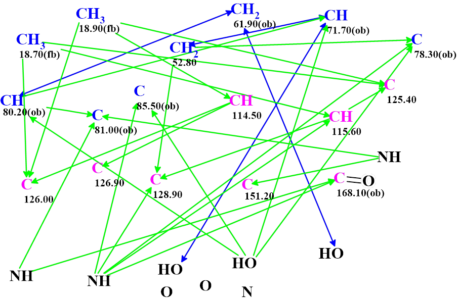
Figure 2. Molecular connectivity diagram (MCD) of mantidisflavin A. Hybridizations of carbon atoms are marked by corresponding colors: sp2 – violet, sp3 – blue. Labels “ob” and “fb” are set by the program to carbon atoms for which neighboring with heteroatom is either obligatory (ob) or forbidden (fb). HMBC connectivities are marked by green arrows and COSY connectivities by blue arrows. The evident C=O bond was drawn manually to accelerate structure generation.
The properties of some atoms have been specified by the user according to known spectral-structural correlations. The presence of a strong carbonyl band in the IR spectrum made it possible manually define a double bond to oxygen from the carbon at 168.10 ppm.
Checking the MCD for the presence of non-standard correlations ended with the following program message: Current Molecular Connectivity Diagram (MCD) passed all tests. No updates performed.
As we see, the program could not detect the presence of a nonstandard correlation. Problem solving statistics showed that the program does not detect the presence of a nonstandard correlation in approximately 10% of cases. This is because of the heuristic algorithm used for analyzing the MCD for the presence of contradictions.
According to the message, a strict structure generation accompanied with the prediction of 13C chemical shifts was initiated.
Results: k = 1054 → (structural filtering) → 28 → (duplicate removal) → 12, tg = 9s.
After performing the 13C chemical shift prediction for all structures of the output file, the structures were ranked in order of increasing average deviations of the calculated chemical shifts from their experimental values. The ranked file is shown in Figure 3.
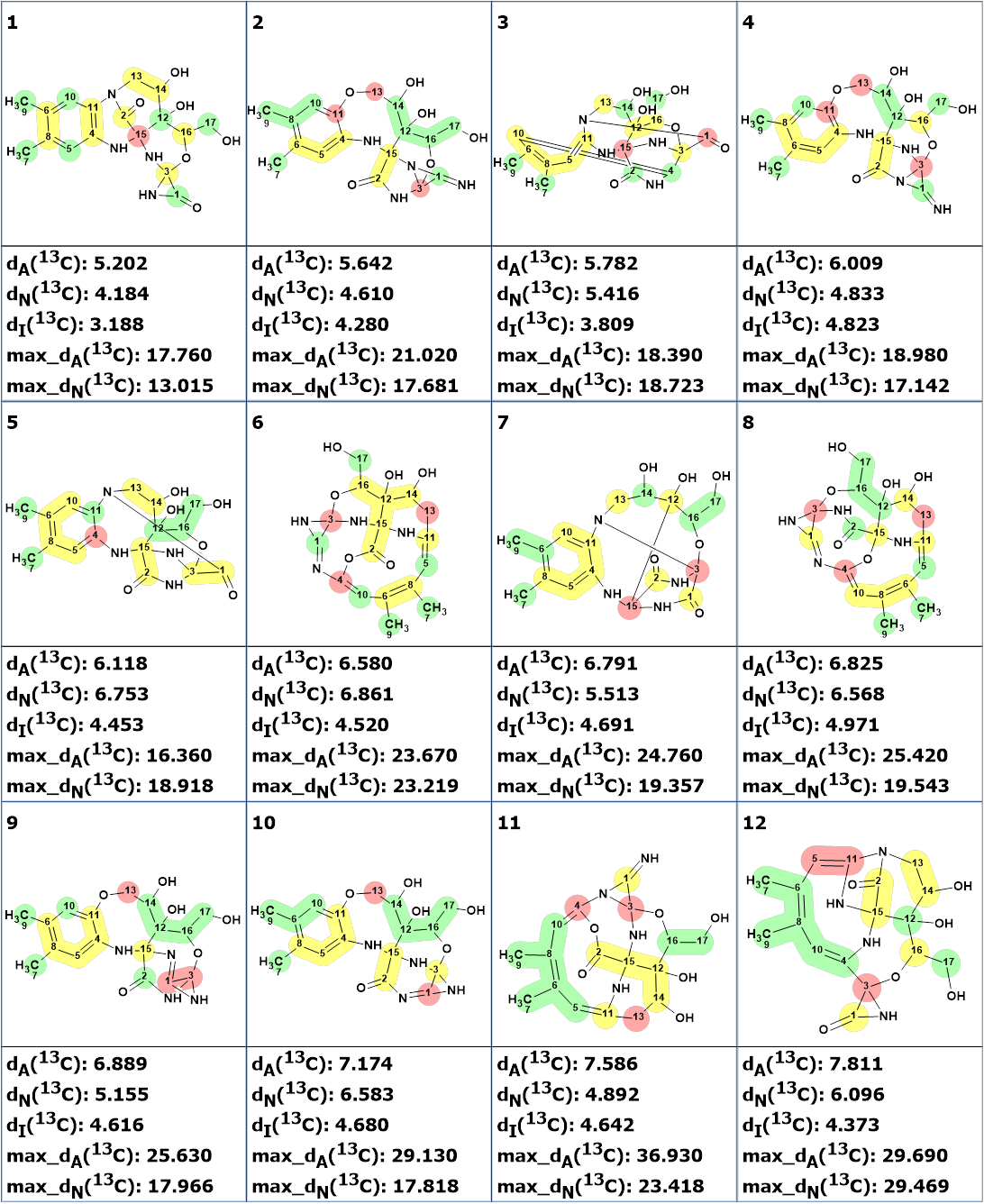
Figure 3. The ranked output file. 13C chemical shift prediction was carried out using the HOSE code-based method, the neural networks, and the incremental approach. Average deviations of 13C chemical shifts determined by these methods are denoted as dA, dN and dI correspondingly. Each atom is colored to mark a difference between its experimental and calculated 13C chemical shifts. The green color represents a difference between 0 to 3 ppm, yellow was >3 to 15 ppm, red > 15 ppm.
Obviously, none of these structures can be correct: the value of the average and maximum deviations is very large, and many structures contain fragments that are not observed in organic molecules let alone natural products. This suggests that there is at least one non-standard correlation in the two-dimensional NMR data. In this case, it is necessary to use fuzzy structure generation [2], which is how the presence of non-standard correlations is detected.
Fuzzy structure generation was launched. The generation options required for the presence of one nonstandard correlation of unknown length.
Result: k = 40,269 → (structural filtering) → 482 → (duplicate removal) → 176,tg=5m 42s. The six top ranked structures are shown in Figure 4.
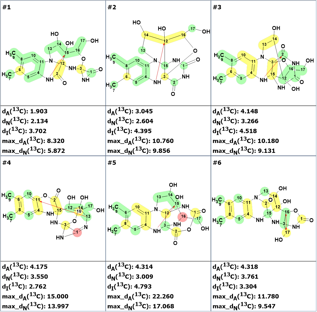
Figure 4. The six top ranked structures of the output file. Ranking is carried out in increasing order of dA deviations.
The minimal deviations are calculated for structure #1. It has one nonstandard correlation (marked by red arrow) which is directed to atom 12 ( but not to atom 15 as in compound 1). However, this molecule contains a fragment improbable in organic chemistry, the three-membered ring with the nitrogen atom. It turned out that neither the ACD/Labs database nor the PubChem library included any structure that contained this fragment.
Structure #2 is identical to the structure of mantidisflavin A determined in [1]. The non-standard correlation in it is the same as the one shown in Figure 1. Structure #4 contains also an uncommon fragment and a correlation of the improbable length of six bonds, while structures #5 and #6 are characterized by large deviations. Therefore, only structures 2 and 3 can be considered as probable structures. The calculation of the empirical DP4 probabilities for these two structures showed that structure #2 does indeed have a high degree of probability (Figure 5).

Figure 5. DP4 probabilities calculated for structures #2 and #3.
Thus, the use of fuzzy structure generation and other options provided in the program made it possible to correctly determine the structure of mantidisflavin A. The 13C chemical shift assignment is shown below:

References
- S.-G. Peng, X.-N. Liu, M. B. Sura, Y.-M. Yan, Y.-X. Cheng. (2024). Mantidisflavin A: A Riboflavin Derivative Featuring a 6/6/6/5/5 Skeleton from the Egg Cases of the Insect Tenodera sinensis Saussure and Its Anti-Renal Fibrosis Activity, Org. Lett., 26, 1316−1320
- M.E. Elyashberg, A.J. Williams. Computer-based Structure Elucidation from Spectral Data. The Art of Solving Problems. Springer, Heidelberg, 2015, 454 p. http://www.springer.com/978-3-662-46401-4
About the Author
