February 28, 2025
by Mikhail Elyashberg, Leading Researcher, ACD/Labs
Setosol
Okeke et al. [1] reported the isolation of a novel catechol, which they named setosol (1), from the fungus Pleiochaeta setosa. Its molecular formula of C15H16O5 was determined from the ESI-MS spectrum.
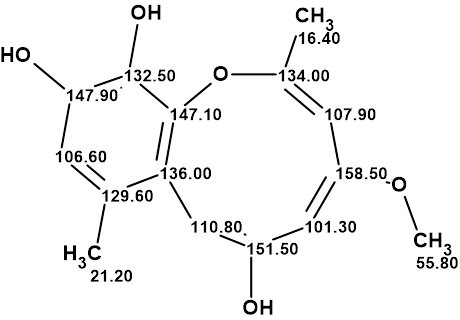
1
Okeke et al. acknowledged that this structure was unique amongst natural products at that time. Indeed, such a ring system also appears to be unknown amongst synthetic compounds. In addition to its unprecedented ring system, setosol is also proposed to be an enol ether. Setosol was subsequently reported to be active against several fungal and bacterial strains. This compound was reisolated from Preussia isomera by Chen et al. who confirmed its anti-bacterial activity [2]. Setosol 1 has also been mentioned in a review without any further comment [3].
Having in mind the unusual properties mentioned of structure 1, Bates et al [4] performed its verification and subsequent revision to structure 2.
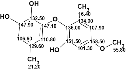
2
To verify structure 1, it was entered into Structure Elucidator Suite and the 13C chemical shifts were predicted using the incremental approach. The results are presented in Figure 1.
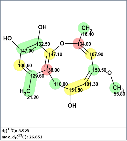
Figure 1. Results of 13C chemical shift prediction for the proposed structure of setosol. Each atom is colored to mark a difference between its experimental and calculated 13C chemical shifts. The green color represents a difference between 0 to 3 ppm, yellow >3 to 15 ppm, and red >15 ppm. The average deviation of predicted chemical shifts from experimental is denoted as dI(13C). A maximum deviation is designated as max_dI(13C).
The large dI(13C) and max_dI(13C) values clearly show that the proposed structure of setosol is incorrect. Though the authors[1] wrote that they used 1H – 13C 2D NMR correlations for the structure elucidation, unfortunately, this information is absent from the article. If 2D NMR data were available as constraints, Structure Elucidator could be used for structure generation in standard mode. Therefore, to reveal a correct structure in a manageable timeframe, a molecular connectivity diagram containing the left (green) part of the benzene ring was created, leaving all the other atoms “free”, so all possible combinations can be explored.

Figure 2. The molecular connectivity diagram (MCD) of setosol. Hybridizations of carbon atoms are marked by corresponding colors: sp2 – violet, sp3 – blue. Labels “ob” and “fb” are set to carbon atoms for which neighboring with heteroatom is either obligatory (ob) or forbidden (fb).
The structure generation was completed in 6 h 20 min, and 8,143,488 structures were produced from which 5783 remained after filtering and duplicate removal. The 13C NMR chemical shift predictions were performed for structures of the output file, and structures were ranked in increasing order of the average deviation value. The twelve top ranked structures are shown in Figure 3.


Figure 3. The twelve top ranked structures of the output file. 13C NMR chemical shift prediction was carried out using the HOSE code-based method, the neural networks, and the incremental approach. Average deviations of 13C NMR chemical shifts determined by these methods are denoted as dA, dN and dI correspondingly. Each atom is colored to mark a difference between its experimental and calculated 13C NMR chemical shifts. The green color represents a difference between 0 to 3 ppm, yellow was >3 to 15 ppm, red > 15 ppm.
The top ranked structure #1 is compound 2, and its validity was confirmed by empirical DP4 probability calculations (see Figure 4) as well as by 13C chemical shift prediction using DFT based approach DU8 ML.
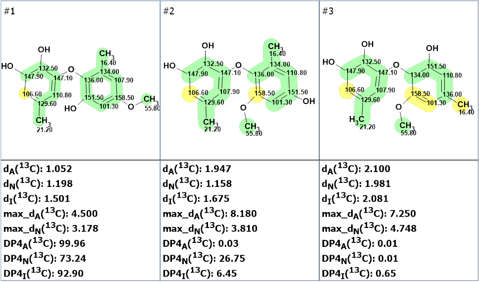
Figure 4. The three top ranked structures of the output file. The 13C NMR chemical shift prediction was carried out using the HOSE code-based method, the neural networks, and the incremental approach. Average deviations of 13C NMR chemical shifts determined by these methods are denoted as dA, dN and dI correspondingly. Each atom is colored to mark a difference between its experimental and calculated 13C NMR chemical shifts. The green color represents a difference between 0 to 3 ppm, yellow was >3 to 15 ppm, red > 15 ppm. DP4A(13C), DP4N(13C) and DP4I(13C) are the probability values that a given structure is correct.
It turned out that structure 2 corresponded to a known natural product, a biaryl ether, which was isolated earlier by several groups. Its structure was reliably established, and biological activity was studied. Comparison of NMR spectra of setosol with those corresponding to structure 2 confirmed the structure revision performed in the work. [1]
References
- B. Okeke, M. Kaouadji, F. Seigle-Murandi, R. Steiman. (1994). Biosci, Biotech, Biochem., 58, 734-736.
- H.-L. Chen, W.-T. Zhao, Q.-P. Liu, H.-Y. Chen, W. Zhao, D.-F. Yang, X.-L. Yang. (2020). Fitoterapia, 141, 104475.
- Agudo-Jurado, F.; Reveglia, P.; Rubiales, D.; Evidente, A.; Barilli, E. (2023). Int. J. Mol. Sci., 24, 5116.
- R. W. Bates, M. Elyashberg, A. G. Kutateladze, C. M. Williams. (2024). Eur. J. Org. Chem. e202400431. https://doi.org/10.1002/ejoc.202400431
About the Author
