

July 21, 2026
by Mikhail Elyashberg, Leading Researcher, ACD/Labs
Computer Assisted Structure Elucidation of Distahlianin A
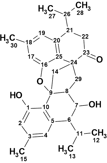
Cadinane-type sesquiterpenoid dimers are structurally rare natural products, with only a limited number having been isolated from higher plants to date. Stahlianthus involucratus (“Wan Gai Pie” in Dai medicine), a traditional medicinal herb native to the Xishuangbanna Dai Autonomous Prefecture of Yunnan, China, has long been used for the treatment of traumatic injuries, rheumatic pain, and related ailments. Previous phytochemical studies have shown that extracts of S. involucratus are rich in cadinane sesquiterpenoids, which exhibit a range of biological activities, including anti-inflammatory, analgesic, antipyretic, and antioxidant effects. A phytochemical investigation by Ji et al. [1] resulted in the isolation of three architecturally unprecedented cadinane sesquiterpenoid dimers, designated distahlianins A–C. Among them, distahlianin A (1) possesses a particularly remarkable carbon skeleton featuring an unusual 5,6-spirocyclic core fused to an oxygenated heterocycle, giving rise to an intricate 6/6/5/6/6/6 hexacyclic framework.
1
The structure of Distahlianin A was determined from the HR-ESI-MS, 1D and 2D NMR, and IR spectra and was confirmed by X-ray analysis. We used this data to challenge the ACD/Structure Elucidator expert system.
Distahlianin A (1) was isolated as colorless crystals from methanol. The HR-ESI-MS spectrum displayed an ion peak at m/z 461.2689 [M+H]+ (calcd. 461.2686), corresponding to the molecular formula C30H36O4 with 13 degrees of unsaturation. The 13C and 1H data, as well as selected HMBC and COSY correlations available from [1] are presented in Table 1.
Table 1. NMR spectroscopic data of Distahlianin A.
| Label | dC | dC calc (HOSE) | XHn | dH | H Mult. | COSY | H to C HMBC |
| C 1 | 157.5 | 153.44 | C | ||||
| C 2 | 116.6 | 114.41 | CH | 6.62 | s | C 10, C 1 | |
| C 3 | 139 | 136.38 | C | ||||
| C 4 | 123.2 | 121.56 | CH | 6.51 | s | ||
| C 5 | 141.8 | 137.31 | C | ||||
| C 6 | 53.8 | 49.75 | CH | 2.7 | t | 2.30, 3.70 | C 10, C 4, C 5 |
| C 7 | 75.5 | 72.88 | CH | 3.7 | u | 2.70, 2.81, 4.13 | |
| C 8 | 49.4 | 52.37 | CH | 2.81 | u | 1.95, 3.70 | C 14, C 9 |
| C 9 | 87.9 | 86.38 | C | ||||
| C 10 | 121 | 121.74 | C | ||||
| C 11 | 27.4 | 28.33 | CH | 2.3 | u | 0.68, 1.13, 2.70 | |
| C 12 | 25.4 | 25.04 | CH3 | 1.13 | d | 2.3 | |
| C 13 | 22.0 | 21.64 | CH3 | 0.68 | d | 2.3 | |
| C 14 | 38.5 | 44.32 | CH2 | 2.19 | u | ||
| C 14 | 38.5 | 44.32 | CH2 | 3 | u | C 29, C 24, C 9, C 25 | |
| C 15 | 21.1 | 21.29 | CH3 | 2.24 | s | C 2, C 4, C 3 | |
| C 16 | 153.9 | 152.79 | C | ||||
| C 17 | 115.7 | 114.29 | CH | 6.47 | s | C 25, C 16 | |
| C 18 | 138.1 | 135.63 | C | ||||
| C 19 | 120.4 | 122.37 | CH | 6.64 | s | C 25 | |
| C 20 | 137.8 | 138.33 | C | ||||
| C 21 | 45.5 | 48.36 | CH | 3.2 | u | 2.16, 2.48 | C 19, C 25, C 20 |
| C 22 | 39.3 | 38.97 | CH2 | 2.88 | u | ||
| C 22 | 39.3 | 38.97 | CH2 | 2.48 | u | 3.2 | C 24, C 23 |
| C 23 | 211.5 | 209.19 | C | ||||
| C 24 | 52.8 | 54.58 | C | ||||
| C 25 | 125 | 120.06 | C | ||||
| C 26 | 33.4 | 32.49 | CH | 2.16 | u | 0.73, 1.02, 3.20 | |
| C 27 | 21.4 | 21.23 | CH3 | 1.02 | d | 2.16 | |
| C 28 | 17.3 | 20.33 | CH3 | 0.73 | d | 2.16 | |
| C 29 | 49.5 | 39.3 | CH2 | 1.95 | u | 2.81 | C 24, C 7, C 9, C 23 |
| C 29 | 49.5 | 39.3 | CH2 | 2.56 | u | ||
| C 30 | 21.3 | 21.13 | CH3 | 2.25 | s | C 17, C 19, C 18 | |
| O 1 | OH | 7.78 | u | C 2, C 10, C 1 | |||
| O 2 | OH | 4.13 | u | 3.7 |
The spectroscopic information shown in Table 1 along with the molecular formula of the compound was entered into Structure Elucidator, and the program created a Molecular Connectivity Diagram, MCD (Figure 1).
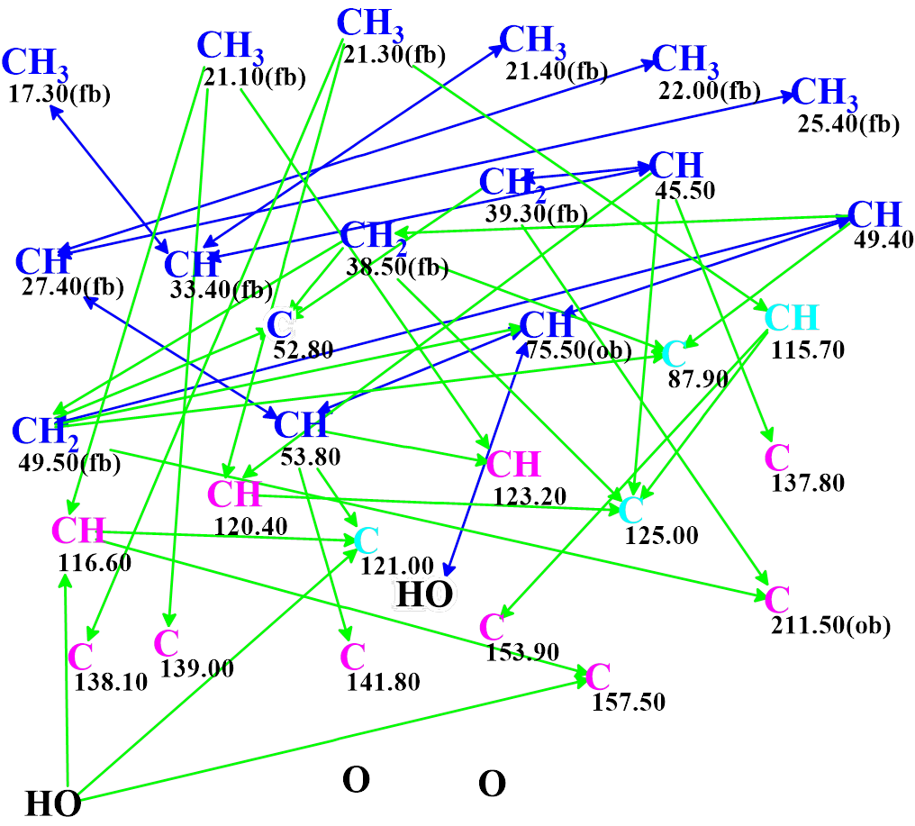
Figure 1. Molecular connectivity diagram (MCD) of distahlianin A. Hybridizations of carbon atoms are marked by corresponding colors: sp2 – violet, sp3 – blue, not sp – light blue. Labels “ob” and “fb” are automatically set by the program to carbon atoms for which neighboring with heteroatom is either obligatory (ob) or forbidden (fb). The HMBC connectivities are marked by green arrows, while COSY connectivities by blue arrows.
We see that MCD contains four light blue carbon atoms with ambiguous hybridization. Consequently, both possible hybridizations (sp3 and sp2) must be checked for all these carbons during the structure generation. Structure generation accompanied by 13C chemical shift prediction and structural filtering was initiated. It was completed with the following result: k = 46 → (Structure Filtering) → 3, tg = 9 s. The three structures of the output file ranked in increasing order of average deviations of calculated chemical shifts from experimental ones are presented in Figure 2.
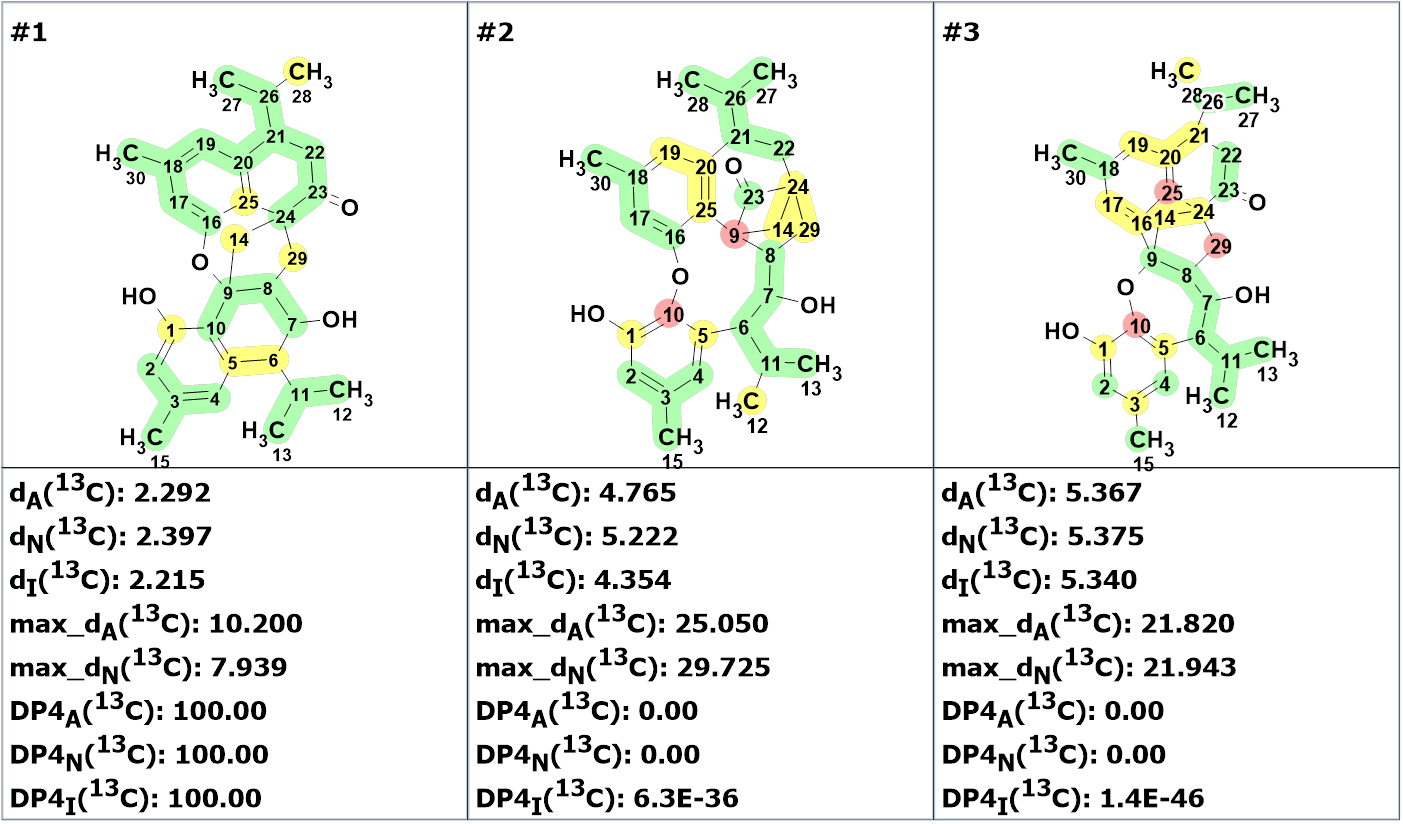
Figure 2. The ranked output file. 13C chemical shift prediction was carried out using the HOSE code-based method, the neural networks, and the incremental approach. Average deviations of 13C chemical shifts determined by these methods are denoted as dA, dN and dI correspondingly. Each atom is colored to mark a difference between its experimental and calculated 13C chemical shifts. The green color represents a difference between 0 to 3 ppm, yellow was >3 to 15 ppm, red > 15 ppm. DP4A, DP4N and DP4I are the probabilities of structure correctness calculated by the program.
Figure 2 shows that the top ranked structure #1 is identical to the structure of distahlianin A shown by the authors of [1]. The calculation of DP4 probabilities was carried out which gave values 100% for all three methods (Figure 2). This allows one to conclude that 13C chemical shift prediction distinguished structure #1 as a correct one with probability of 100%.
In the work [1], only key HMBC and COSY correlations were graphically represented on the molecular structure. Authors of many publications assume that such a connectivity representation confirms the correctness of the derived structure. Figure 3 shows the structures generated with the HMBC and COSY correlations visualized with arrows.
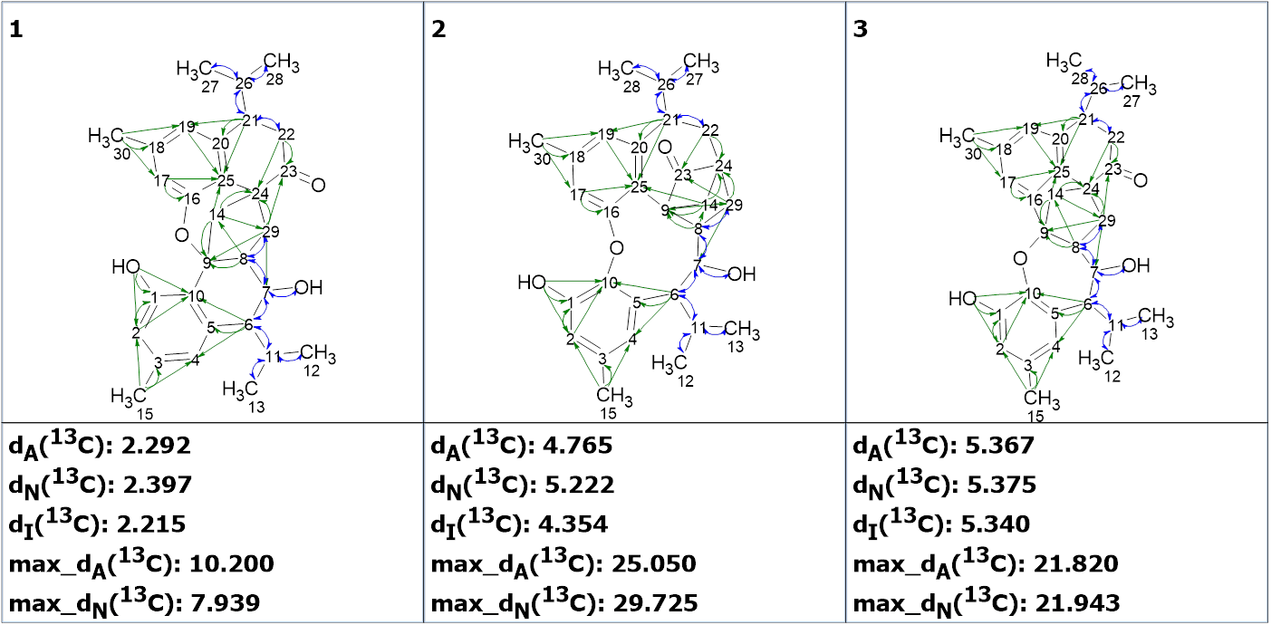
Figure 3. Structures of the output file. HMBC and COSY correlations are shown by green and blue arrows correspondently.
We see that all structures fully correspond to the key correlations, and the correct structure is revealed only using the prediction of chemical shifts. The incorrect structures have large average and maximum deviations. Therefore, a graphical representation of the correlations on a structure does not prove its correctness but only indicates that the given structure is consistent with the 2D NMR spectral data.
In conclusion, the structure of a new natural product characterized by a structurally unique carbon skeleton containing an intricate 6/6/5/6/6/6 hexacyclic framework was unambiguously and reliably elucidated in a few seconds using Structure Elucidator in fully automatic mode.
References
- K.-L. Ji, Y.-H. Chen, Q.-Q. Song, X.-Y. Zheng, P. Sun, J.-M. Lu, G. Liang, Y.-Y. Fan. (2025). Distahlianins A−C: Architecturally Diverse Cadinane-Type Sesquiterpenoid Dimers from Stahlianthus involucratus. Org. Lett., 27, 10843−10848.
About the Author