August 1, 2013
by Mikhail Elyashberg, Leading Researcher, ACD/Labs
Asidia SAAF
Sperm activation and chemotaxis occur ubiquitously in animal species, including humans, and play an important role in fertilization. To understand the molecular mechanism underlying the genus-specificity of sperm chemotaxis of ascidians (a type of marine invertebrate filter feeders), Matsumori et al [1] have investigated the structure of a novel compound Ascidia-SAAF 2 (1) isolated from the eggs of the ascidian Ascidia sydneiensis.
1
Ascidia-SAAF 2 was isolated from HPLC fractions in the amount of 4 nmol (2.6 µg). The chemical structure of Ascidia-SAAF 2 was elucidated by performing NMR spectroscopy and mass spectrometry. The molecular formula of Ascidia-SAAF 2, C27H44O10S2Na2, was obtained from the negative-ion high-resolution MS (m/z 296.1188, [M-2Na]2-, calculated m/z 296.1193), indicating a dehydrogenated or oxygenated (-H2) form of the known compound Ciona-SAAF 1 (C27H46O10S2Na2).
The gDQF-COSY and gHMBC spectra obtained using cold-probe technology unambiguously demonstrated the presence of a double bond between C-22 and C-23. Although chemical shifts of H 22 and H 23 almost overlapped to provide second-order signals, its E configuration was identified on the basis of a large 3JHH (17 Hz) between H 22 and H 23, deduced from a spectral simulation of the signals. The absence of ROE between H 21 and H 24 could lend support for the presence of this configuration.
The spectroscopic NMR data used for the molecular structure elucidation with ACD/Structure Elucidator are presented in Table 1.
Table 1. Ascidia-SAAF 2. Spectroscopic NMR data.
| Label | δC | δC calc | CHn | δH | M(J) | COSY | C HMBC |
| C 1 | 33.3 | 37.13 | CH2 | 1.18 | u | 1.90, 3.56 | |
| C 1 | 33.3 | 37.13 | CH2 | 1.55 | u | 1.84 | |
| C 2 | 26.5 | 26.54 | CH2 | 1.8 | u | ||
| C 2 | 26.5 | 26.54 | CH2 | 1.84 | u | 1.55, 4.65 | |
| C 3 | 78.5 | 81.9 | CH | 4.65 | u | 1.60, 1.84 | |
| C 4 | 18 | 34.16 | CH2 | 1.6 | u | 1.90, 4.65 | |
| C 4 | 18 | 34.16 | CH2 | 1.55 | u | ||
| C 5 | 32.6 | 39.37 | CH | 1.9 | u | 1.18, 1.60 | |
| C 6 | 31.5 | 32.04 | CH2 | 1.9 | u | ||
| C 6 | 31.5 | 32.04 | CH2 | 1.18 | u | ||
| C 7 | 72 | 72.4 | CH | 3.56 | u | 1.18 | |
| C 8 | n.d. | 76.14 | C | ||||
| C 9 | 50.2 | 51.14 | CH | 1.25 | u | 1.59 | |
| C 10 | 36 | 37.68 | C | ||||
| C 11 | 32.9 | 20.04 | CH2 | 1.59 | u | 1.21, 1.25 | |
| C 12 | 40.7 | 37.06 | CH2 | 1.21 | u | 1.59 | |
| C 12 | 40.7 | 37.06 | CH2 | 1.99 | u | ||
| C 13 | 43.2 | 42.42 | C | ||||
| C 14 | 53.9 | 54.35 | CH | 1.54 | u | 1.38 | |
| C 15 | 18.6 | 24.3 | CH2 | 1.47 | u | ||
| C 15 | 18.6 | 24.3 | CH2 | 1.38 | u | 1.28, 1.54 | |
| C 16 | 28.4 | 28.43 | CH2 | 1.28 | u | 1.14, 1.38 | |
| C 16 | 28.4 | 28.43 | CH2 | 1.73 | u | ||
| C 17 | 56.6 | 55.04 | CH | 1.14 | u | 2.07, 1.28 | |
| C 18 | 13.5 | 20.57 | CH3 | 0.9 | u | C 13, C 12, C 9, C 17, C 14, C 1, C 10, C 5 | |
| C 19 | 11.5 | 21.58 | CH3 | 0.9 | u | ||
| C 20 | 39.7 | 32.67 | CH | 2.07 | u | 0.98, 1.14, 5.39 | |
| C 21 | 20.3 | 20.85 | CH3 | 0.98 | u | 2.07 | C 22, C 20, C 17 |
| C 22 | 140.5 | 140.59 | CH | 5.39 | u | 2.02, 2.07 | |
| C 23 | 125.5 | 128.38 | CH | 5.39 | u | ||
| C 24 | 36 | 33.32 | CH2 | 1.93 | u | C 22, C 23 | |
| C 24 | 36 | 33.32 | CH2 | 2.02 | u | 5.39, 1.87 | |
| C 25 | 33.5 | 34.07 | CH | 1.87 | u | 0.92, 2.02, 3.83 | |
| C 26 | 74.1 | 76.27 | CH2 | 3.83 | u | 1.87 | |
| C 26 | 74.1 | 76.27 | CH2 | 3.95 | u | ||
| C 27 | 16.4 | 17.41 | CH3 | 0.92 | u | 1.87 | C 26, C 24, C 25 |
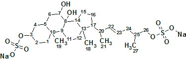
The Molecular Connectivity Diagram from which the structure generation was initiated is presented in Figure 1.
Figure 1. Ascidia-SAAF 2. Molecular Connectivity Diagram.
MCD overview. Two SO4Na fragments were drawn by hand as their presence was postulated by authors [1] from previous chemical knowledge. A signal of one carbon atom was not observed in the 13C NMR spectrum, so it was introduced in Table 1 without any chemical shift (C 8). This atom is most probably a quaternary one and it was marked as “sp2 or sp3“-hybridized on the MCD (light blue). It is seen from Table 1 (italic underlined) that there are three pairs of overlapped 1H signals and this is due to the many ambiguous COSY and HMBC connectivities (dotted lines). At the same time, all sp3-hybridized carbon atoms are supplied with labels “fb” or “ob” defining their admissible neighbors. These postulated atom properties accelerate the structure generation significantly and reduce size of the output structural file in spite of the plethora of ambiguous connectivities.
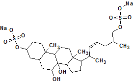
No contradictions were detected in 2D NMR data, and the Strict Structure Generation combined with 13C chemical shift prediction (using the incremental method) was initiated with an average deviation threshold (d=4 ppm). The results were: k=2880→20→8, tg = 31.5 s (including the time of spectrum prediction). The two top structures of the ranked output file are presented in Figure 2.
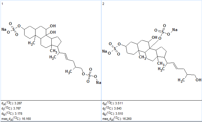
Figure 2. Ascidia-SAAF 2. The two top structures of the ranked output
file. The double bond is in E configuration.
Figure 2 shows that the top-ranked structure coincides with the structure of Ascidia-SAAF 2; though the difference Δ=dA(2)-dA(1) is small, the individual deviation values are reasonably large, which is possibly explained by the presence of SO4Na fragments. Table 1 shows that values of experimental and predicted 13C chemical shifts of atoms C 3 and C 26 (which are neighbors of SO4Na in structure #1 and #2 respectively) are very similar.
As seen in Table 1, the largest discrepancies between experimental and calculated chemical shifts are observed for atoms C4 and C11. Chemical Shift Calculation Protocols were generated for these atoms and are presented in Figures 3 and 4.
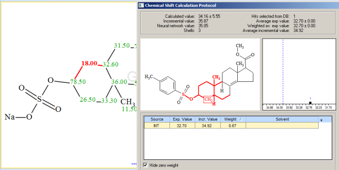
Figure 3. Ascidia-SAAF 2. Chemical Shift Calculation Protocol for the carbon atom C 4 (18.00 ppm).
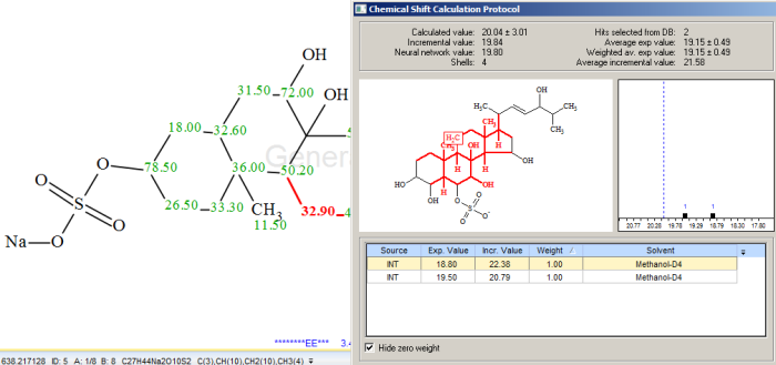
Figure 4. Ascidia-SAAF 2. Chemical Shift Calculation Protocol for the carbon atom C 11 (32.90 ppm).
Only one hit from the internal database was found in the first case, and two hits in the second, which is why the calculated chemical shifts do not closely match the experimental data. We also noticed that the calculated shift for C 4 (34.11) more closely matches the experimental chemical shift for C 11, and vice versa, which indicates that the assignments of the experimental shifts (18.00 and 32.90 ppm) should be exchanged for these two atoms. When the assignments were exchanged as shown below, the average deviations for structure #1 reduced to the following values: dA=2.278, dI=2.864 and dN=2.170 ppm.
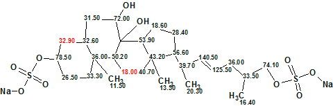
Structure #1, highlighting the exchanged C 4 and C 11 chemical shift assignments.
We believe that this result can be justifies the exchange of chemical shift assignments.
The single double bond C=C exists in structure #1 in E configuration which was established by authors [1] on the basis of a large 3JHH (17 Hz) between H 22 and H 23, deduced from a spectral simulation of the signals and on the ROESY data (see above). However 13C chemical shift prediction can also help in distinguishing between E and Z configurations. When structure #1 was transferred to Z configuration, all deviations increased significantly (to dA=2.584, dI=3.079 and dN=2.422 ppm), confirming that the E configuration is correct.
Structure #1, Z configuration
Therefore the correct structure was determined and its configuration upon a double bond was able to be identified by only the aids of Structure Elucidator. The 13C chemical shift of the carbon C 8 that was not observed in the spectrum is predicted to be 76 ppm.
References
- N. Matsumori, Y. Hiradate, H. Shibata, T.Oishi, S. Shimma, M.Toyoda, F. Hayashi, M. Yoshida, M.Murata, M. Morisawa. A Novel Sperm-Activating and Attracting Factor from the Ascidian Ascidia Sydneiensis. Org. Lett., 2013, 15 (2), pp 294–297.
About the Author
