February 1, 2013
by Mikhail Elyashberg, Leading Researcher, ACD/Labs
Asperjinone
Liao et al1 isolated and elucidated the structure of a new natural product named as Asperjinone (1) presented in Figure 1. This compound was isolated, along with other 12 known compounds, from Aspergillus terreus. Our analysis using ACD/Structure Elucidator resulted in the revision of structure 1; we suggested that structure 2 is the correct structure (see Figure 1). The story of the structure revision is described in the recent article in the Journal of Natural Products.2
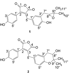
Figure 1. The original structure of Asperjinone (1) and the revised structure, 2.
The molecular formula C22H20O6 and the NMR data presented in Table 1 were obtained from the original work1 and used as input into the Structure Elucidator software.
Table 1. Asperjinone. 1D and 2D Spectroscopic data (600 MHz, Acetone-d6).
| Position | δC | Type | δH (J in Hz) | HMBCa |
|---|---|---|---|---|
| 1 | 165.7 | C | ||
| 2 | 140.7 | C | ||
| 3 | 137.5 | C | ||
| 4 | 166.8 | C | ||
| 5 | 29.2 | CH2 | 3.97, d (11.2)3.98, d (11.2) | C-2, 3, 4, 1″, 2″ |
| 1′ | 119.0 | C | ||
| 2′, 6′ | 131.5 | CH | 7.63, d (8.1) | C-2, 1′, 2′, 4′ |
| 3′, 5′ | 115.8 | CH | 7.01, d (8.1) | C-1′, 4′ |
| 4′ | 160.3 | C | ||
| 1″ | 127.5 | C | ||
| 2″ | 129.6 | CH | 6.99, m | C-4″, 6″, 7″ |
| 3″ | 120.9 | C | ||
| 4″ | 152.2 | C | ||
| 5″ | 117.0 | CH | 6.66, d (8.6) | C-3″, 4″ |
| 6″ | 127.3 | CH | 6.99, m | C-5 |
| 7″ | 31.2 | CH2 | 2.67, dd (16.9, 8.0) 2.94, dd (16.9, 5.0) | C-2″, 3″, 4″, 8″, 9″ |
| 8″ | 68.8 | CH | 3.76, m | |
| 9″ | 77.0 | C | ||
| 10″ | 19.7 | CH3 | 1.22, s | C-8″, 9″, 11″ |
| 11″ | 25.3 | CH3 | 1.33, s | C-8″, 9″, 10″ |
aHMBC correlations, optimized for 6 Hz, are from the proton(s)
stated to the indicated carbon.
MCD overview. The Molecular Connectivity Diagram (MCD) created automatically by the program is presented in Figure 2. Two atoms (colored in pale blue) in the MCD—C(119.0) and C(120.9)—were classified as having ambiguous hybridization because these chemical shifts are characteristic for both the C=C double bonds (sp2) and for C(sp3) atom if it is included into an O-C-O fragment. Carbons with chemical shifts falling into the interval 152–167 ppm are likely connected with at least one oxygen atom. The information presented in the MCD was used by the program for the purpose of structure generation. As a result all structures proposed by the software were in agreement with the HMBC correlations and atom properties, and no additional manual modifications based on common HMBC correlations were necessary. No structural inputs regarding the presence of aromatic rings or other conceivable rings in the structure were made.
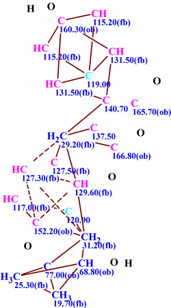
Figure 2. Asperjinone. The Molecular Connectivity Diagram (MCD) extracted from the spectroscopic data. Atoms are artificially arranged in such a manner which approximately corresponds to atom positions in revised structure 2.
The structure generation process produced the following results: k=3658→2641→1939, tg = 1m 50s. This indicates that 3658 isomeric structures were generated in 1m 50s, and 2641 structures were stored after spectral and structural filtering. 13C NMR chemical shifts were then calculated for the stored structures using an incremental approach (this procedure took 8 sec), and duplicate structures were removed to give 1939 structures. The output structural file was ranked in ascending order of the chemical shift deviation, then 13C chemical shifts were predicted for all 1939 structures using a neural network based program (14 seconds calculation time) and then for the first 15 structures of the ranked file using a HOSE code based program (1 minute calculation time). The first 9 structures of the ranked file are displayed in Figure 3. Atoms for which the Δ = |δCcalc – δCexp| value (the difference between experimental and calculated chemical shifts) is less than 3 ppm are marked in green; yellow correspond to D=3–15 ppm and red to D>15 ppm. The figure shows that the first ranked structure (all green) is characterized by the smallest deviations calculated by HOSE code and neural network based methods, while the structure proposed by Liao and co-workers1 was placed in third position by the ranking procedure. The deviation is almost twice the size of that given for the structure ranked in first position.
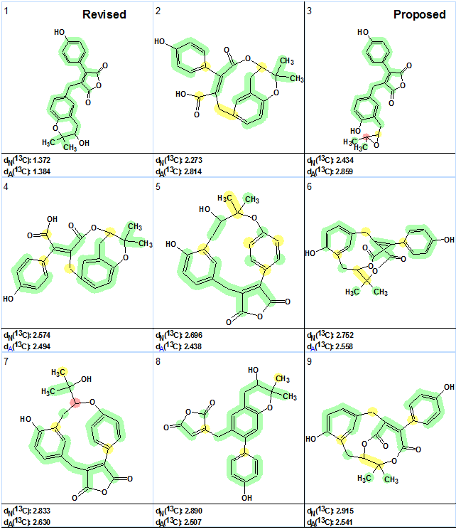
Figure 3. Asperjinone. The first 9 structures of the output file ranked by deviations calculated using a neural network and HOSE code based 13C NMR prediction programs. Colored circles on the atoms display chemical shift differences. Green color denotes a difference less than 3 ppm, yellow—between 3 and 15 ppm, and red—more than 15 ppm. Designation of deviations: dA—HOSE code based algorithm, dN—neural network based algorithm.
To confirm the revised structure, 2, we performed a search for the (3,3-dimethyloxiran-2-yl)methyl fragment, as shown below and with the numbering system included for easy recognition of the substructure in the reference molecule.
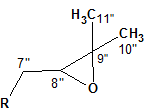
Figure 4. (3,3-dimethyloxiran-2-yl)methyl fragment
A fragment search was carried out in the ACD/NMR Database (included with Structure Elucidator) containing 425,000 structures with assigned 13C and 1H chemical shifts. The program selected almost 180 structures from which approximately 150 structures were chosen that exhibit the closest similarity with the environment of the oxirane fragment. For these structures, a scatter plot was created (see Figure 4). Here the 13C chemical shifts related to the C-8″ and C-9″ atoms of structure 1 are presented for all selected structures. The chemical shift values (69 and 77 ppm) assigned to the corresponding atoms C-8″ and C-9″ in the original structure 1 are also shown by their labels on the right side of the graph.
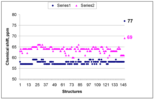
Figure 5. Asperjinone. A scatter plot of the 13C chemical shift values related to atoms 8″ and 9″ of the original structure 1. Series 1 (blue circles) corresponds to atom 9″ (dC 77 ppm in structure 1), series 2 (pink triangles) – to atom 8″ (dC 69 ppm in structure 1).
Inspection of the scatter plot convincingly confirms the incorrectness of the original structure: the chemical shifts of C-8″(68.8 ppm in structure 1) are observed in the range of 60-65 ppm while for C-9″(77.0 ppm in structure 1) the corresponding range is 57-59 ppm.
On the other hand, corroboration of the revised structure 2 was found in the Supporting Information of the original work1. One of the compounds separated by the authors1 along with Asperjinone (designated as Butyrolactone V) was characterized and its 13C and 1H NMR chemical shifts were assigned to the structure of Butyrolactone V. This compound contains the revised structural component of structure 2. Both structures supplied with the assigned 13C chemical shifts (for butyrolactone V only partial assignment is shown) are presented in Figure 5.
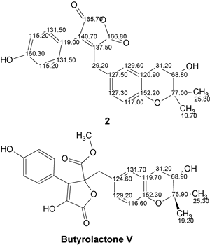
Figure 6. Comparison of chemical shifts in the revised part of structure 2 with those in Butyrolactone V.
The comparison leaves no doubts regarding the correctness of structure 2. Moreover, oxirane 1JCH couplings are typically ~180 Hz, far larger than other oxygen-bearing aliphatic carbon. If the authors1 had measured this value for the C8″ atom they would easily realize that their suggested structure was wrong.
Therefore, the suggestion of the authors1 regarding the existence of an oxirane ring in the Asperjinone structure proved to be erroneous. We believe that the application of a CASE system to the structure elucidation of this natural product would have allowed the authors to avoid this incorrect structure as an output from their analysis.
It should be noted that as far as we know this is the first example when reliable structure revision was performed only with the aid of CASE system without performing additional experiments or quantum chemical NMR shift calculations.
References
- Liao, W.-Y.; Shen, C.-N.; Lin, L.-H.; Yang, Y.-L.; Han, H.-Y.; Chen, J.-W.; Kuo, S.-C.; Wu, S.-H.; Liaw, C.-C.; Asperjinone, a Nor-Neolignan, and Terrein, a Suppressor of ABCG2—Expressing Breast Cancer Cells, from Thermophilic Aspergillus terreus. J. Nat. Prod., 75:630-635, 2012.
- Elyashberg, M.E., Blinov K.A., Molodtsov S.G., Williams A.J. Structure Revision of Asperjinone Using Computer-Assisted Structure Elucidation Methods. J. Nat. Prod, 2013, ASAP, DOI: 10.1021/np300218g
About the Author
